
Abbas Dhami
Specialist Diagnostic Radiographer
Knee pain is one of the most common reasons people visit their GP in the UK, and knee osteoarthritis is one of the leading causes of long-term disability in adults over fifty. Yet for all the appointments, scans, and treatments that patients go through, a fundamental question often goes unanswered: why is this knee wearing down faster than it should? In the vast majority of cases, the answer lies not just in the joint itself, but in how the entire leg is aligned and standard imaging almost never checks this properly.
The way your knee is loaded every time you stand, walk, or climb stairs depends entirely on how straight and balanced your lower limb is from hip to ankle. When that alignment is even slightly off, one side of the knee absorbs far more pressure than the other, and over years that imbalance grinds cartilage away unevenly, accelerating joint damage. This is precisely where EOS scan knee imaging changes the conversation.
By capturing the full lower limb in a natural standing position — with your body weight exactly as it is in real life — EOS imaging for knee alignment gives clinicians a level of structural detail that no lying-down scan can provide, and a foundation for knee arthritis treatment planning that is grounded in how your body actually works.
What Is Knee Osteoarthritis and Why Does Alignment Matter?
Knee osteoarthritis is far more than a simple wear-and-tear condition — its progression is directly shaped by how forces move through the joint, which means alignment plays a central role in both its cause and its treatment.
What Happens Inside an Arthritic Knee
Knee osteoarthritis occurs when the protective cartilage cushioning the ends of the bones inside the joint gradually breaks down. As cartilage thins, the space between the bones narrows, the joint loses its smooth movement, and bone begins to rub against bone. The result is pain, stiffness, swelling, and over time a progressive loss of function. It develops slowly over many years and is more common on one side of the knee than the other — a pattern that itself points toward uneven loading rather than simple ageing.
Why Knee Alignment Is the Missing Piece
The knee sits between the hip and the ankle, and its long-term health depends entirely on the straightness of that chain. When knee alignment is off even by a few degrees, body weight concentrates on one compartment of the joint. Over thousands of steps per day, this uneven pressure accelerates cartilage loss in the overloaded area, often decades before the other side shows any damage.
Without understanding and correcting that alignment, even the best treatments address the damage without ever changing the conditions that created it.
Why Standard Knee Scans Often Miss the Full Picture
Most patients receive their first knee scan lying flat on a bed — which is precisely the position in which the key mechanical problems are least likely to show up.
The Limits of Lying-Down Imaging
When you lie down for an MRI or standard knee X-ray, body weight is no longer travelling through the joint. The knee opens slightly, muscles relax, and the alignment the leg adopts in this position is entirely different from the one it holds during daily activity.
Any misalignment that is only significant under load — which is often the case with early and mid-stage knee osteoarthritis — can look unremarkable on a lying-down scan. Clinicians may find cartilage damage but have no way to assess the mechanical reason it appeared, or why it concentrates on one side.
What a Weight-Bearing Knee X-Ray Reveals That Others Cannot
A weight-bearing knee X-ray or standing knee X-ray captures the joint under exactly the conditions it experiences in real life. In this position, joint space narrowing is real and functional — not an artefact of a relaxed, unloaded position. The degree of varus or valgus misalignment — the technical terms for bow-legged and knock-kneed positions — can be assessed accurately, and the actual mechanical stress on each compartment becomes visible.
This is a fundamentally more useful piece of diagnostic information than any lying-down scan can provide. Before your scan, it is worth reviewing the eligibility criteria at ScanAlign to understand whether a weight-bearing EOS knee assessment is the right next step for you.
What Is EOS Imaging and How Does It Work for the Knee?
EOS imaging takes the principle of weight-bearing imaging several steps further — producing a level of detail, precision, and whole-body context that no conventional standing X-ray can match.
The Technology Behind the EOS Scan
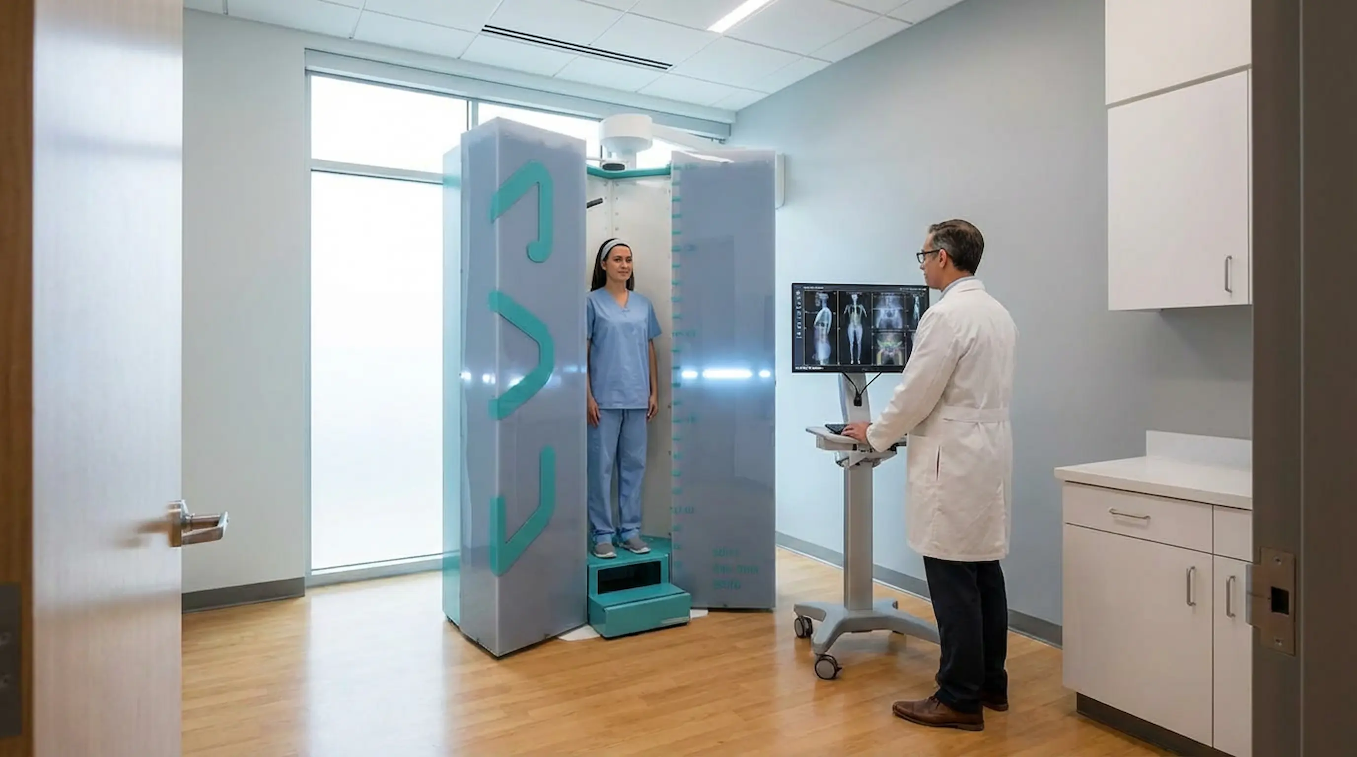
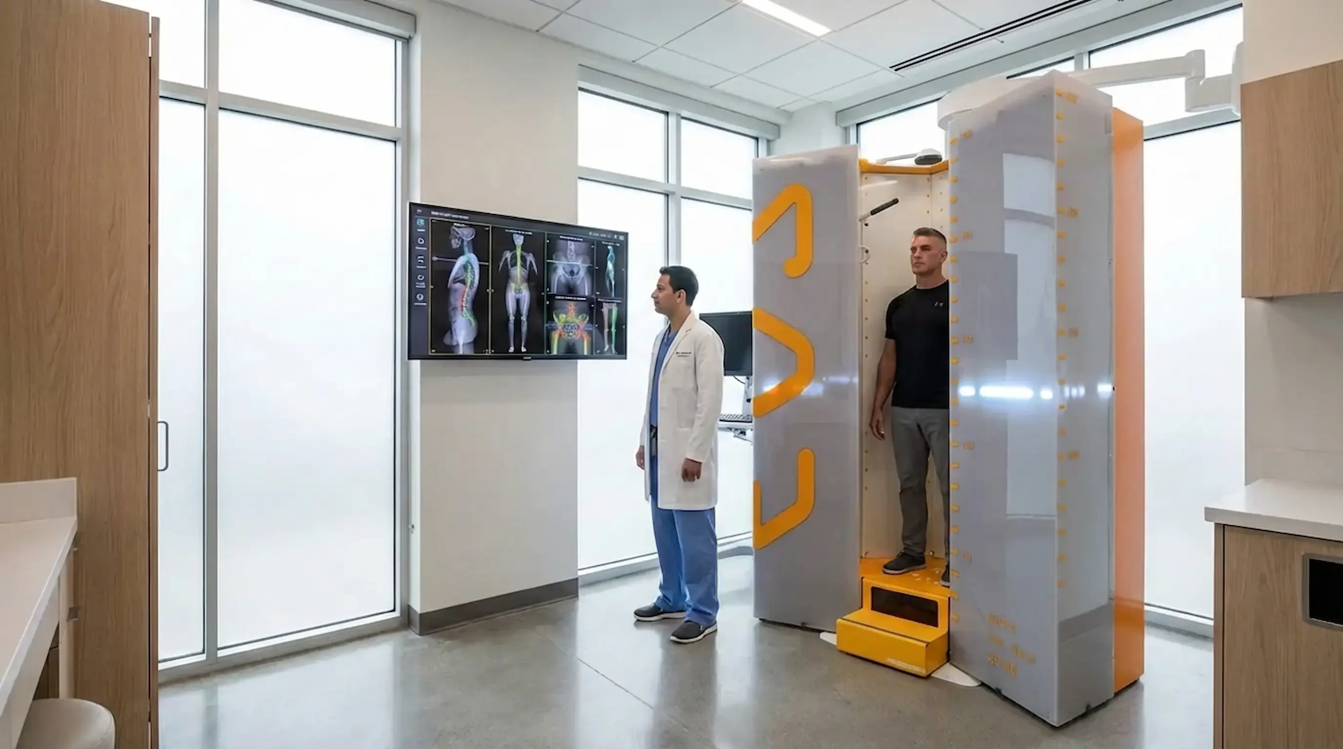
EOS imaging is a full-body scanning system that captures simultaneous front and side images of your entire skeleton while you stand in a natural, relaxed position. For knee assessment, this means the hip, knee, and ankle are all imaged at the same moment in the same scan, with no repositioning and no image stitching.
The system uses Nobel Prize-winning particle detector technology, originally developed at CERN, which produces sharp, clinically precise images at up to 90% less radiation than a standard X-ray.
EOS Imaging for Knee Alignment: The Full Lower Limb in One Shot
Unlike any other imaging method, EOS imaging for knee alignment shows the entire lower limb in correct 1:1 scale while you stand under full body weight, revealing every factor that determines how much pressure your knee is carrying and where it concentrates:
- A conventional X-ray shows only the knee; EOS shows how the knee sits within the whole skeleton
- Whether the hip is tilted and shifting load unevenly down through the joint
- Whether one leg is shorter and forcing the knee to compensate with every step
- Whether the ankle is rotating inward and altering the mechanical axis of the entire limb
What Does an EOS Scan Reveal About Your Knee?
A single EOS scan knee produces clinical data that goes far beyond what conventional imaging can offer, giving specialists both the detail of the joint and the structural context of the whole lower limb.
Knee Alignment and the Mechanical Axis
The most important measurement for understanding knee osteoarthritis progression is the hip-knee-ankle angle — the line running from the centre of the hip joint, through the knee, and down to the ankle. In a well-aligned leg, this line passes through the centre of the knee. When knee alignment is off, it shifts to one side, concentrating load on the inner or outer compartment. EOS imaging for knee alignment measures this angle precisely in a standing, weight-bearing position — quantifying exactly how far off centre the load is sitting, which no lying-down scan can achieve.
EOS Long Leg Imaging and Leg Length Discrepancy
Even a small difference in leg length meaningfully affects how the knee is loaded. When one leg is shorter, the pelvis tilts to compensate, shifting the mechanical axis of the longer leg and increasing stress on one side of the knee. EOS long leg imaging captures both legs in a single standing scan, measuring their true length under body weight and showing precisely how any discrepancy is affecting knee alignment and load distribution.
Grading Knee Osteoarthritis Severity With EOS
Research confirms that EOS imaging for knee osteoarthritis is as accurate as conventional radiography for grading arthritis severity using the Kellgren-Lawrence scoring system. Joint space width, bone changes, and cartilage loss can all be assessed reliably from EOS images — and because measurements are taken in a standing position, they reflect the actual functional state of the joint rather than a decompressed, resting version of it.
EOS Imaging vs. MRI and Standard X-Ray for Knee Arthritis
Each imaging method reveals something different, and understanding those differences is essential for choosing the right diagnostic path for your knee condition.
The table below outlines how EOS imaging, MRI, and a standard knee X-ray compare across the key areas that matter for knee osteoarthritis treatment planning:
| Feature | EOS Imaging | Standard X-Ray | MRI |
|---|---|---|---|
| Patient position | Standing, weight-bearing | Usually lying or partial load | Lying flat |
| View | Hip to ankle in one scan | Knee only | Knee only |
| Mechanical axis measurement | Precise | Limited | Not available |
| Leg length assessment | Yes | No | No |
| Soft tissue detail | Limited | Limited | Excellent |
| Radiation dose | Very low | Low | None |
When EOS and MRI Work Better Together
EOS and MRI are not competitors — they answer different clinical questions. EOS imaging maps bones, alignment, and the mechanical forces acting on the knee under real load. MRI examines the soft tissue consequences: cartilage damage, ligament stress, and inflammation. When both are used together, clinicians gain the fullest possible picture: the structural cause from EOS, and the tissue-level impact from MRI. For patients managing a complex knee osteoarthritis treatment pathway or approaching surgery, this combination significantly improves decision-making quality.
How EOS Supports Knee Arthritis Treatment Planning
Accurate imaging does not just describe a problem — it fundamentally changes the quality of every treatment decision that follows, from rehabilitation through to surgery.
Non-Surgical Treatment: Orthotics, Braces, and Physiotherapy
The precise measurements from an EOS scan for knee alignment give physiotherapists and orthotists real, weight-bearing data about how the knee is loaded and where the alignment is failing. For orthotics and knee braces, a device designed using standing EOS measurements fits the patient’s actual mechanics, making it far more effective at redistributing load away from the damaged compartment than one based on estimated averages.
EOS Surgical Planning: Preparing for Knee Replacement
For patients approaching knee replacement or corrective surgery, accurate pre-operative alignment data is one of the most important factors in determining long-term outcomes. 3D knee planning EOS tools allow surgeons to work from a precise, patient-specific model of the lower limb, assessing bone angles, planning implant size and position, and simulating different surgical approaches before making a single incision.
Monitoring Recovery After Knee Surgery
EOS imaging is used post-operatively to confirm the implant has restored the correct mechanical axis, to check healing progress, and to identify any compensatory changes developing elsewhere such as the hip or lower back adjusting to a newly aligned knee. Because EOS uses such low radiation, repeat scans during recovery are safe and practical, providing objective data at every stage rather than relying on symptoms alone.
Signs Your Knee Pain Could Be an Alignment Problem
Not all knee pain has the same origin — and certain symptom patterns point specifically toward a structural alignment issue rather than isolated joint damage.
Common Symptoms That Point to Misalignment
Knee pain does not always feel the same, and the specific pattern of your symptoms can reveal a great deal about whether a structural alignment problem is at the root of it. If any of the following sound familiar, a standing X-ray for knee arthritis or EOS imaging for knee arthritis may finally explain why your pain keeps returning despite treatment:
- Pain that is consistently worse on one side of the knee rather than spread evenly across the joint
- Pain that appears specifically when climbing stairs, standing for long periods, or walking downhill
- Intermittent knee pain that comes and goes depending on activity level
- Recurring soft tissue injuries around the knee, including sprains, tendon problems, or repeated muscle strain
How EOS Confirms What Symptoms Only Suggest
Symptoms are signals — but they are not measurements. No amount of clinical history-taking can quantify the degree of varus misalignment or tell a surgeon exactly how far the mechanical axis is displaced from centre. Only EOS imaging for knee alignment delivers that level of precision — turning a pattern of symptoms into a specific set of structural findings that can be directly and confidently acted upon.
If you are unsure whether your symptoms match the profile for an EOS knee scan, the patient support team at ScanAlign can help you make that decision before you commit to an appointment.
EOS Imaging for Knee Osteoarthritis: Who Should Consider It?
EOS is a genuinely useful diagnostic tool for a much broader range of patients than those already facing surgery, and access to it is more straightforward than most people expect.
Patients Who Benefit Most
You are likely to benefit from an EOS knee scan if you:
- Have knee pain that is consistently worse on one side of the joint
- Have been told your scans are normal but symptoms continue to worsen
- Carry a diagnosis of knee osteoarthritis and want to understand its structural cause
- Are an athlete with recurring knee injuries or alignment-related pain
- Are preparing for knee replacement surgery and want to support pre-operative planning
- Are recovering from knee surgery and want to confirm alignment has been restored
- Have noticed that one leg appears slightly shorter or that your knees angle inward or outward
Why Low-Dose Imaging Matters for Long-Term Monitoring
Managing knee osteoarthritis requires ongoing assessment as the condition evolves. Because weight-bearing EOS imaging delivers up to 90% less radiation than a standard X-ray, repeat scanning over months or years is safe and practical.
ScanAlign also offers a dedicated paediatric EOS imaging service for younger patients with developing lower limb conditions. For a clear explanation of what EOS radiation exposure actually means for your health, visit is an EOS scan safe on the ScanAlign website.
Your Step-by-Step Guide to Getting an EOS Knee Scan at ScanAlign
Getting an EOS knee scan at ScanAlign is a clear, structured process built entirely around giving you precise answers and a direct path to better care.
Step 1 — Book a Free Video Consultation
Start with a free video consultation with an EOS specialist. Your symptoms, medical history, and any previous imaging are reviewed, and the specialist confirms whether a knee EOS scan is the right approach for your situation.
Step 2 — Attend Your Scan at Harley Street
Your scan takes place at ScanAlign’s Harley Street clinic. You stand inside the open EOS scanning cabinet in a natural, comfortable position while front and side images of your complete lower limb and skeleton are captured in around 15 seconds — no lying down, no discomfort, no claustrophobia.
Step 3 — Receive Your Expert Radiology Reports
UK-trained radiology specialists analyse your images and produce detailed expert reports covering your knee alignment, mechanical axis measurements, leg length data, and any structural findings, giving your surgeon, physiotherapist, or orthotist an immediate, actionable foundation.
Step 4 — Review Your Results With a Doctor
A doctor walks you through every finding in plain, clear language. You receive a GP referral summary to support ongoing care, and your full rights throughout the process including access to your complete results are protected in line with the patients' rights policy at ScanAlign. To hear what the experience is like from patients who have already been through it, visit the ScanAlign patient feedback page before booking.
Conclusion
Knee osteoarthritis is not simply a matter of a joint wearing out — it is almost always a story of misalignment, uneven loading, and structural forces acting on the joint for years without ever being properly measured. EOS imaging changes that, delivering precise, weight-bearing, full lower-limb data that no lying-down scan can replicate, and giving clinicians the foundation they need to treat the actual mechanical cause rather than just managing the damage it leaves behind.
Compared to traditional imaging, EOS shows how your body functions under natural weight. It is quick, painless, and accurate — helping doctors identify the real cause of knee pain, posture issues, or joint problems and plan better treatment.
Book Your EOS Full Body Scan Today →FAQs
- What does an EOS scan show for knee osteoarthritis? It shows the full lower limb alignment — hip, knee, and ankle — under body weight, measuring the mechanical axis, varus or valgus misalignment, leg length discrepancy, and osteoarthritis severity in one standing scan.
- How is EOS imaging different from a standard knee X-ray? A standard X-ray images only the knee, usually lying down. EOS images the entire lower limb in two views simultaneously while you stand under full body weight, revealing the mechanical cause of knee problems, not just the damage.
- Can EOS imaging help plan knee replacement surgery? Yes. EOS surgical planning tools provide a precise 3D model of the lower limb, allowing surgeons to assess bone angles and plan implant position before the operation, which can improve alignment accuracy.
- Is a weight-bearing EOS knee scan safe to repeat regularly? Yes. EOS uses up to 90% less radiation than a conventional X-ray, making it one of the safest options for long-term repeat monitoring of knee osteoarthritis.
- Who should have an EOS scan for knee alignment? Anyone with one-sided knee pain, a knee osteoarthritis diagnosis, recurring knee injuries, visible leg misalignment, or upcoming knee surgery may benefit. Check the eligibility criteria at scanalign.co.uk.
Continue reading
More articles
Is EOS Scan Safe? Radiation Levels, Risks, and FAQs
Discover if EOS imaging is safe, its radiation levels, risks, and benefits. Learn why it’s preferred for accurate, low-dose skeletal scans.

EOS Scan for Scoliosis: Why Standing Imaging Matters
Discover how EOS for scoliosis provides low-radiation imaging for accurate spinal alignment and better scoliosis diagnosis and monitoring.
Hip Pain & Alignment: EOS Imaging for Better Diagnosis
EOS imaging for hip pain reveals pelvic tilt, leg length discrepancy & spinopelvic alignment that routine scans miss. Book Appointment now.