
Abbas Dhami
Specialist Diagnostic Radiographer
You did everything right. Went to the doctor, described the pain honestly, got referred for an MRI, and waited. The results came back and you were told it looks fine, nothing serious was found. But fine is not how your back feels when getting out of bed each morning, after sitting at a desk for a few hours, or by the end of a long day on your feet. The pain is still there, and now you have no explanation for it.
Here is something that rarely gets said: a clear MRI does not mean nothing is wrong with you. It means the scan did not find anything in the areas it was designed to look at, taken in the position it was taken. Spinal alignment issues, pelvic tilt, and postural imbalances are a whole separate category of pain causes that standard scans are simply not built to detect. For many people living with unexplained back pain, that is exactly where the real answer has been hiding all along.
What Does "MRI Shows Nothing" Actually Mean?
An MRI is a useful scan for discs, soft tissue, and nerve detail, but it is always performed lying flat, completely still, with your body fully unloaded. Your muscles relax, your pelvis settles, and the spine decompresses from the weight it carries all day. That is not the position your body is in when the pain actually happens, and that one fact changes everything about what the scan can and cannot find.
What MRI does well:
- Detecting disc herniations, bulges, and degeneration
- Showing nerve compression and spinal canal narrowing
- Identifying soft tissue damage and inflammation
- Spotting tumours, infections, and serious structural problems
What MRI cannot show:
- How your spine and pelvis align when you are standing
- Pelvic tilt angle under real body weight
- Spinal curves as they actually behave in daily life
- Full lower limb alignment and compensation patterns
What Is Spinal Alignment and Why Does It Matter?
Your spine, pelvis, and lower limbs work together as one connected system, constantly balancing and adjusting around each other. When one part shifts out of its natural position, everything above and below it compensates to cope. That slow, ongoing compensation is what quietly turns into daily pain over weeks and months without any obvious injury or damage ever happening.
Why spinal alignment matters:
- Even a small pelvic tilt changes how the entire spine sits above it
- Poor spinal curves increase pressure on lumbar discs and joints
- Misalignment forces muscles on one side to overwork constantly
- Long-term compensation can lead to chronic pain in the back, hips, and knees
- All of this happens while standing and is completely invisible on a lying-down scan
If you want to understand how posture can affect pain more broadly, you can also read ScanAlign’s guide on back pain and poor posture.
How Poor Posture and Pelvic Tilt Cause Pain That Scans Miss
The pelvis sits at the base of the spine, and if it tilts even slightly, everything above it has to adjust around that shift. That adjustment changes the curve of the lower back, moves the hips out of position, and places uneven load through the lumbar vertebrae day after day. Over time, that pressure becomes ongoing pain that feels structural but never shows on a spine scan because the problem is positional, not physical damage.
How pelvic tilt drives widespread pain:
- Compresses the lumbar spine and facet joints unevenly
- Shifts the hip socket position, causing one-sided hip pain
- Changes how load travels through the knee on every step
- Forces the upper spine to compensate, building mid and upper back tension
- Creates tightness on one side and weakness on the other
- Keeps pain coming back after treatment because the root cause is never properly addressed
Anterior vs Posterior Pelvic Tilt: What Is the Difference?
Pelvic tilt comes in two forms, and both can cause real pain. But they pull the body in opposite directions and need different treatment approaches. Understanding which one applies to you starts with measuring it properly.
| Feature | Anterior Pelvic Tilt | Posterior Pelvic Tilt |
|---|---|---|
| What happens | Front of pelvis tips down, back tips up | Front of pelvis tips up, back tips down |
| Lower back effect | Excessive arch in the lower back | Lower back curve flattens out |
| Common cause | Prolonged sitting, tight hip flexors | Standing too long, weak glutes |
| Pain location | Lower back, hip flexors, groin area | Lower back, hamstrings, tailbone |
| Visible sign | Belly pushed forward, exaggerated arch | Flat backside, tucked-under posture |
| Shows on MRI? | No — pelvis shifts when lying flat | No — same reason |
| Shows on EOS? | Yes — precisely measured standing | Yes — precisely measured standing |
Both types cause real daily pain. Both are invisible on a lying-down scan. And both respond better to treatment once properly identified through standing alignment imaging.
Common Signs Your Back Pain Could Be an Alignment Problem
Before any scan, your body often gives you clues that alignment is the real issue. These are the patterns worth paying attention to:
- Pain noticeably worse after standing or walking for 20–30 minutes
- One-sided tightness in the lower back or hip that never fully clears
- Pain eases when lying down but returns quickly once you are upright again
- Uneven hips or shoulders when someone checks your posture from behind
- Recurring back pain that returns after physio or treatment, never fully resolving
- Unexplained hip pain or knee pain on one side with no injury ever found
- A feeling your posture is off, but it has never been properly measured
If several of these sound familiar, alignment deserves a proper investigation, not another lying-down scan.
Important Note: Pelvic tilt and spinal alignment problems do not appear on a standard MRI. When you lie flat, the pelvis naturally adjusts and the spine decompresses, masking the exact imbalances that cause pain when you are standing and moving through your day.
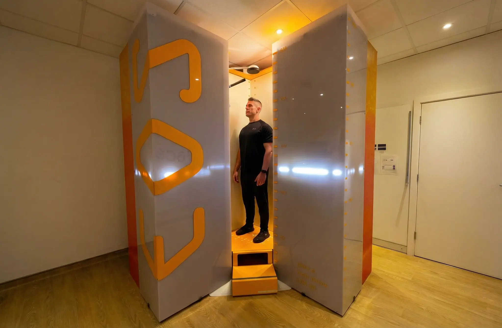
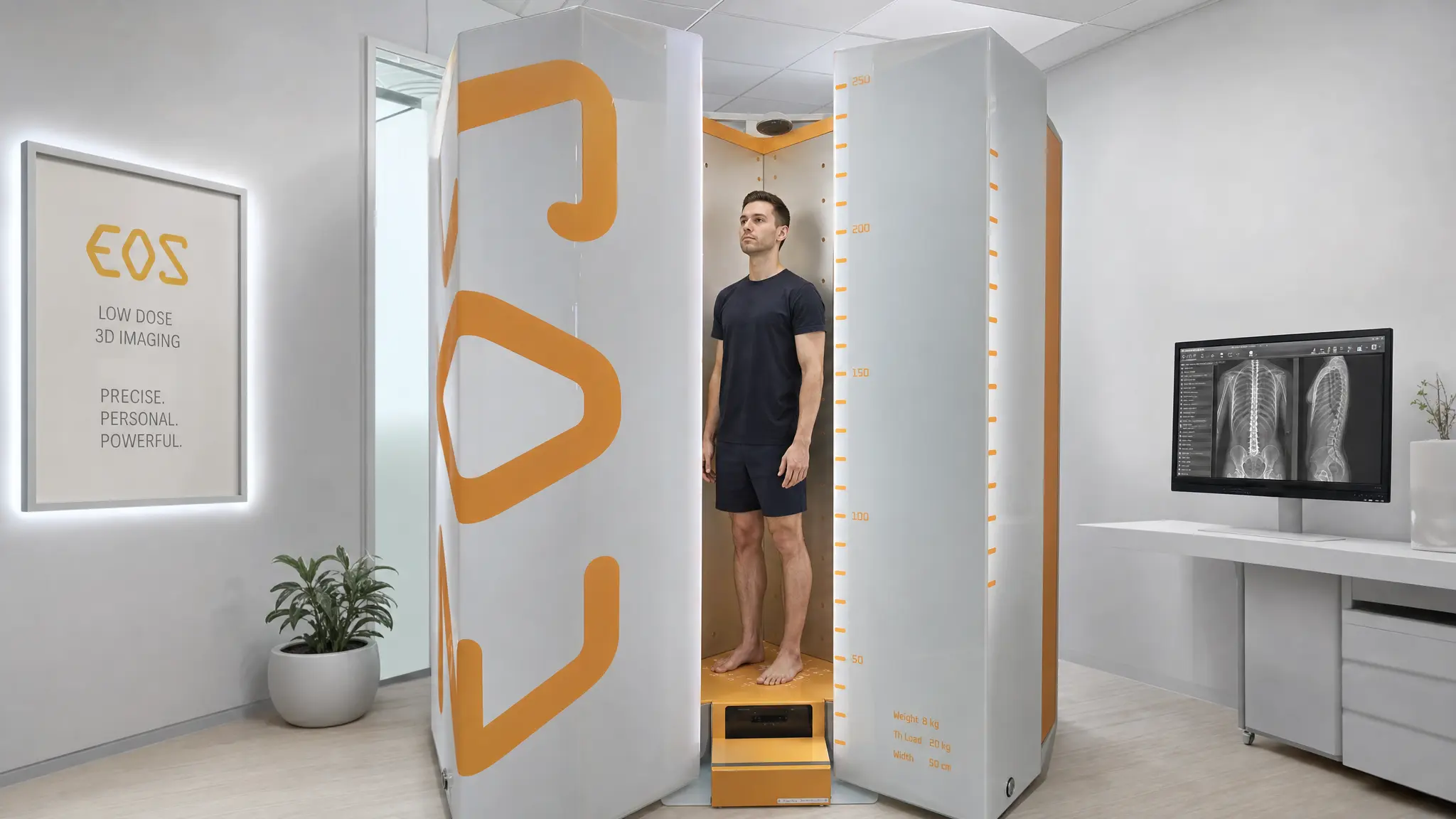
What Is EOS Imaging and How Is It Different?
EOS imaging is a full-body scanning system that works in a completely different way to standard imaging. Instead of lying down, you stand naturally inside a scanning cabin while two X-ray beams move from head to foot simultaneously, capturing a front and side image of your entire skeleton at the same time. The whole scan takes around 15 seconds.
What makes it different is three things working together at once:
- Standing position: Your body is scanned exactly as it functions every day, upright and under full load.
- Full body in one image: Skull to feet in a single continuous scan, not separate sections joined together.
- 3D reconstruction: Both images combine into a three-dimensional skeletal model with precise measurements of every curve, angle, and alignment parameter.
The technology uses Nobel Prize-winning detector technology originally developed at CERN. At ScanAlign, based at Harley Street Hospital in London, it is used every day for patients whose pain has not been explained by standard imaging.
You can also read more about what an EOS scan is and how it works before booking your appointment.
What EOS Imaging Can Reveal That MRI Cannot
This is where EOS imaging for back pain completely changes the picture. While MRI shows tissue and disc detail, EOS shows how your whole skeleton actually sits and loads when you are standing. From a single EOS scan, a consultant radiologist can measure:
- Exact pelvic tilt angle and direction
- Lumbar lordosis — whether the lower back curve is too deep or too flat
- Thoracic kyphosis — the shape and depth of the upper back curve
- Full spinopelvic balance — how the spine and pelvis relate to each other
- Lower limb alignment from hip to ankle on both sides
These are the exact parameters that can drive chronic back pain, hip pain, and knee pain in people whose MRI comes back clear. Weight-bearing imaging gives doctors something a lying-down scan never can: a picture of your body under the actual conditions that create the pain.
EOS vs MRI: A Clear Side-by-Side Comparison
Most people have only ever had an MRI and assume it gives the full picture. It does not, and this table shows exactly where the difference lies between the two scans.
| Feature | EOS Imaging | MRI |
|---|---|---|
| Body position | Standing — weight-bearing | Lying flat — fully unloaded |
| Shows alignment | Full 3D alignment data | No alignment measurement |
| Pelvic tilt measurement | Precise angle and direction | Not possible lying down |
| Full body view | Head to foot, one image | Section by section only |
| 3D reconstruction | Yes | No |
| Radiation | Very low | None, magnetic field |
| Best for | Alignment, posture, balance | Disc, nerve, soft tissue detail |
Both scans have genuine value in the right situation. But for back pain that MRI has not explained, EOS is the scan that looks at the body the way it actually functions.
Who Should Consider EOS Imaging for Back Pain?
Doctors recommend EOS scans across many situations, but it is particularly worth considering if:
- Your MRI came back clear but the back pain has not gone away
- You have hip pain or knee pain on one side with no structural cause found
- You have been told your posture is poor, but it has never been properly measured
- Chronic musculoskeletal pain keeps returning despite physio or treatment
- You are preparing for or recovering from spinal or hip surgery
- You are an athlete with persistent one-sided back, hip, or knee pain
- You can see visible postural imbalance, uneven hips, rounded shoulders, or a lean to one side
You do not need a GP referral at ScanAlign. You can self-refer and get answers without waiting.
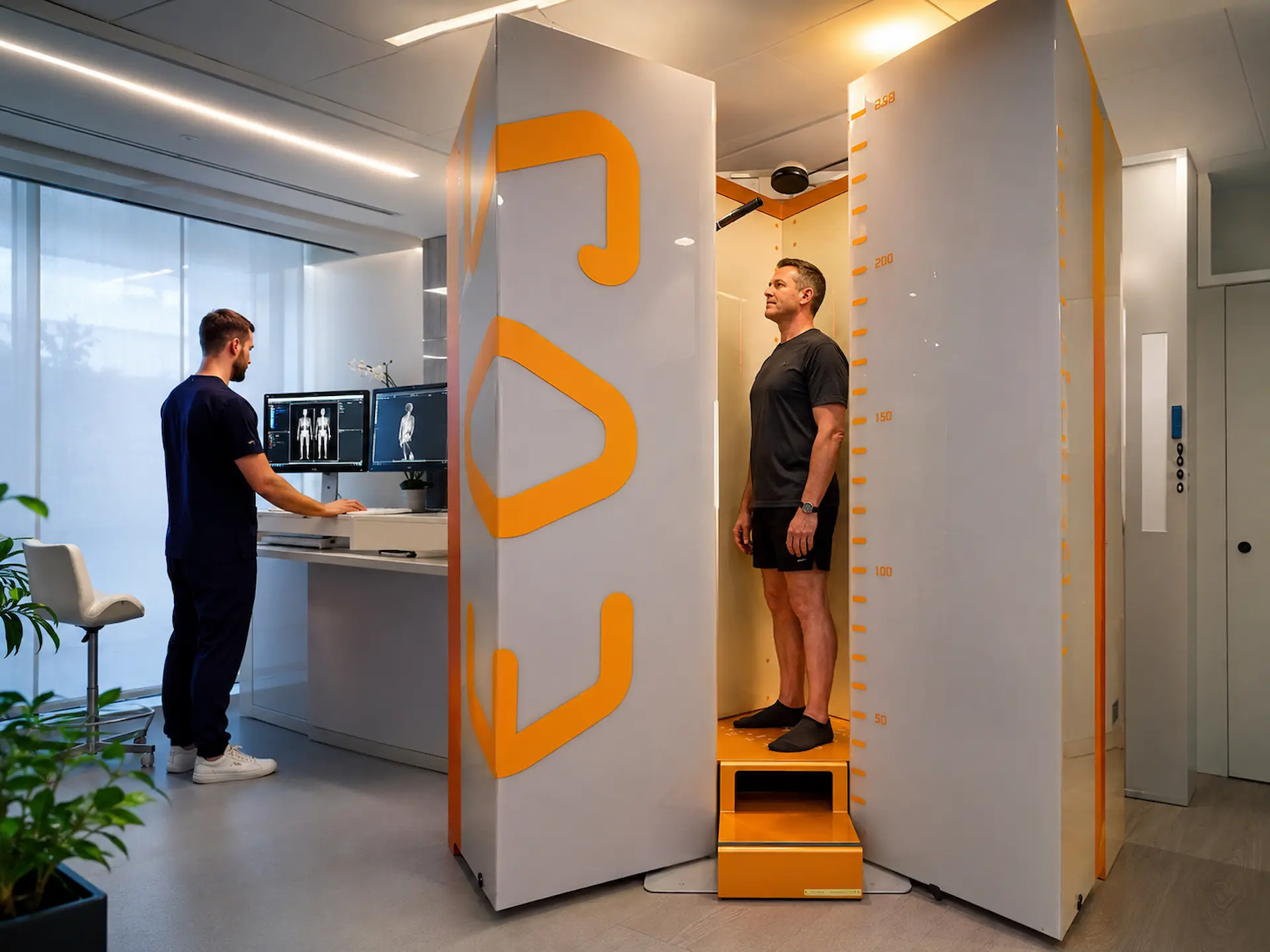
What Happens at a ScanAlign EOS Appointment?
The whole process is simple from start to finish. Here is what to expect step by step:
Free Video Consultation
A specialist reviews your symptoms and history before you travel anywhere, to confirm EOS is the right next step for you.
Arrive at Harley Street Hospital
No injection, no contrast dye, and no preparation are needed. Just wear comfortable clothing and remove any metal items before the scan.
15-Second Standing Scan
You stand naturally in the cabin while the system scans head to toe. There is no tunnel, no tight space, and nothing uncomfortable.
Consultant Radiologist Report
A UK-trained specialist analyses your images and produces a detailed report including your 3D skeletal model and all alignment measurements.
Doctor Follow-Up
A doctor walks through every finding with you, explains what it means, and discusses what treatment options make sense for your situation.
Find out more about what happens during an EOS scan if you want to know exactly what to expect before your appointment.
What Alignment Problems Look Like on an EOS Report
When your EOS report comes back, it gives your doctor precise measurements rather than just images, and that changes the treatment conversation completely. Instead of estimating from a lying-down picture, your clinician can see exactly what your alignment looks like under real body weight.
What the EOS alignment report includes:
- Pelvic tilt angle: Exact degree and direction of any tilt present
- Lumbar lordosis measurement: Whether the lower back curve is normal, too deep, or flattened
- Thoracic kyphosis data: Shape and depth of the upper back curve
- Full spinopelvic balance: How the spine and pelvis relate to each other overall
- 3D skeletal model: Your skeleton shown in your natural standing position
- Lower limb alignment: Side-by-side measurements comparing both sides
For someone whose pelvic tilt is driving their back pain, this is often the first time the actual problem has been measured rather than guessed at. That makes all the difference to what treatment is recommended next.
How Alignment Problems Are Treated Once Found
Finding the alignment problem is step one. What happens next is where the real difference shows. Treatment depends on exactly what the EOS report reveals, but the most common approaches include:
- Targeted physiotherapy working on specific muscles pulling the pelvis or spine out of position
- Posture correction programmes built around exact EOS measurements, not generic exercises
- Orthotics or shoe lifts where a leg length difference is contributing to the tilt
- Surgical planning for cases where the misalignment is structural and significant enough to require it
- Ongoing monitoring for patients who need regular review to track alignment changes over time
The difference from general back pain treatment is significant. Instead of treating broadly and hoping, the clinician treats the specific alignment pattern that is actually causing the pain.
Is EOS Imaging Safe?
Yes, and the safety record is one of the things that genuinely sets EOS apart. The radiation dose is up to 90% lower than a standard X-ray and dramatically lower than a CT scan, so patients do not have to choose between getting useful information and protecting themselves from unnecessary exposure.
Key EOS safety facts:
- Up to 90% less radiation than a conventional X-ray
- Far lower exposure than a CT scan or CT scanogram
- Safe for repeat scanning over months and years of monitoring
- Appropriate for children, adults, and older patients alike
- No injection, no contrast dye, and nothing physically uncomfortable
- Performed at CQC-licensed Harley Street Hospital under consultant radiologist supervision
Read more about EOS scan safety and radiation levels if you want the full detail before booking your appointment.
Important Note: EOS delivers up to 90% less radiation than a standard X-ray, making it one of the safest full skeletal imaging options available, including for patients who need repeat scans over a long period of monitoring.
Conclusion
A normal MRI is not a dead end. It is a gap. Standard scans look at the body lying flat, checking for disc problems and tissue damage. They were never built to assess how your spine, pelvis, and lower limbs align when you are actually standing, loaded, and getting through your day. That gap is real, and it is where a huge amount of chronic back pain actually lives. If your MRI came back clear but the pain never left, alignment has simply not been investigated properly yet.
EOS imaging looks at the body the way it actually functions: standing up, under load, in full 3D. The pain you have been feeling has been real all along. The right scan can finally show exactly why.
You Have Been in Pain Long Enough. Find Out What Is Actually Causing It.
If your MRI came back clear but the pain never left, alignment may be what nobody has checked yet. At ScanAlign, Harley Street Hospital, a 15-second standing EOS scan gives consultant radiologists a complete 3D picture of your spinal alignment, the kind of picture a lying-down scan simply cannot provide. Book your free video consultation today. No referral needed.
FAQs
- 1. Can back pain be caused by alignment even if MRI shows nothing? Yes. MRI scans tissue, discs, and nerves while you lie flat. It does not measure spinal alignment, pelvic tilt, or how your skeleton loads when you are standing. Alignment-driven pain is a separate category that MRI is not designed to pick up.
- 2. What is EOS imaging and how does it work? EOS takes simultaneous front and side X-ray images of your full skeleton while you stand naturally upright. Those two images are combined into a 3D model, giving precise measurements of spinal curves, pelvic tilt, and lower limb alignment.
- 3. Does EOS scan show pelvic tilt and spinal alignment? Yes. This is exactly what EOS was built to do. It measures pelvic tilt angle, lumbar lordosis, thoracic kyphosis, and full spinopelvic balance in a standing position.
- 4. Is EOS imaging better than MRI for diagnosing postural back pain? For postural and alignment-related pain, yes. MRI is better for disc, nerve, and soft tissue detail. EOS is better for understanding how your whole skeleton sits and loads when upright.
- 5. How long does an EOS alignment scan take at ScanAlign? The scan itself takes around 15 seconds. Your full appointment, including preparation and initial discussion, is typically 30–45 minutes. A consultant radiologist report and doctor follow-up are included as part of the process.
- 6. Can poor posture cause chronic back pain and knee or hip pain? Absolutely. Poor posture and pelvic tilt change how load travels through the spine, hips, and knees every day. Over time, this can build uneven pressure and chronic pain across all three areas.
Continue reading
More articles
What Is an EOS Scan?
Learn what an EOS full body scan is, how it works, its benefits, safety, and how it helps detect spine, joint, and posture problems with low radiation imaging.
EOS Scan for Leg Length Discrepancy: Why Accurate Measurement Matters
Hip or back pain with no clear answer? Leg length could be the cause. EOS checks both legs while you stand and finds what standard lying-down scans always miss.
Posture Assessment in London: When Should You Consider an EOS Scan?
Got back pain or posture problems in London? Find out when a posture assessment and EOS scan can help you uncover the real cause of your pain.