
Abbas Dhami
Specialist Diagnostic Radiographer
Athletes often believe that better results come only from training harder, but performance is also affected by how well the body is aligned during movement. Hidden structural problems can quietly change the way an athlete runs, jumps, lands, and absorbs force, leading to recurring injuries or unexplained performance limits. This is where musculoskeletal imaging becomes valuable, as it helps reveal issues beneath the surface that may not be obvious through pain alone. With 3D skeletal imaging for athletes, specialists can now assess alignment while the body is standing in its natural weight-bearing position.
Many athletes spend years managing the same recurring pain without discovering the true cause. In many cases, the problem is not training effort but small alignment imbalances that create stress in joints and muscles over time. These hidden issues can slowly build into serious injuries if left unnoticed. A full-body structural assessment through standing 3D imaging offers a proactive way to identify these risks early, helping athletes improve injury prevention, movement efficiency, and long-term performance.
Why Injury Prevention Needs Better Assessment
Training load gets blamed for most sports injuries. But load is rarely the full picture. Two athletes can follow the exact same programme one stays healthy while the other keeps breaking down. The difference often comes down to how their body is built and how it moves.
Many recurring injuries are connected to movement patterns, poor posture, and misaligned structures that place too much stress on specific areas. When these issues go undetected, the same injury keeps returning no matter how carefully training is managed. Early injury risk assessment shifts the focus from treating the symptom to understanding the cause and for athletes at any level, that awareness is genuinely valuable.
The good news is that modern sports medicine now has better tools for this purpose tools that assess the full body, in a standing position, under real gravitational load.
What 3D Skeletal Imaging Actually Shows
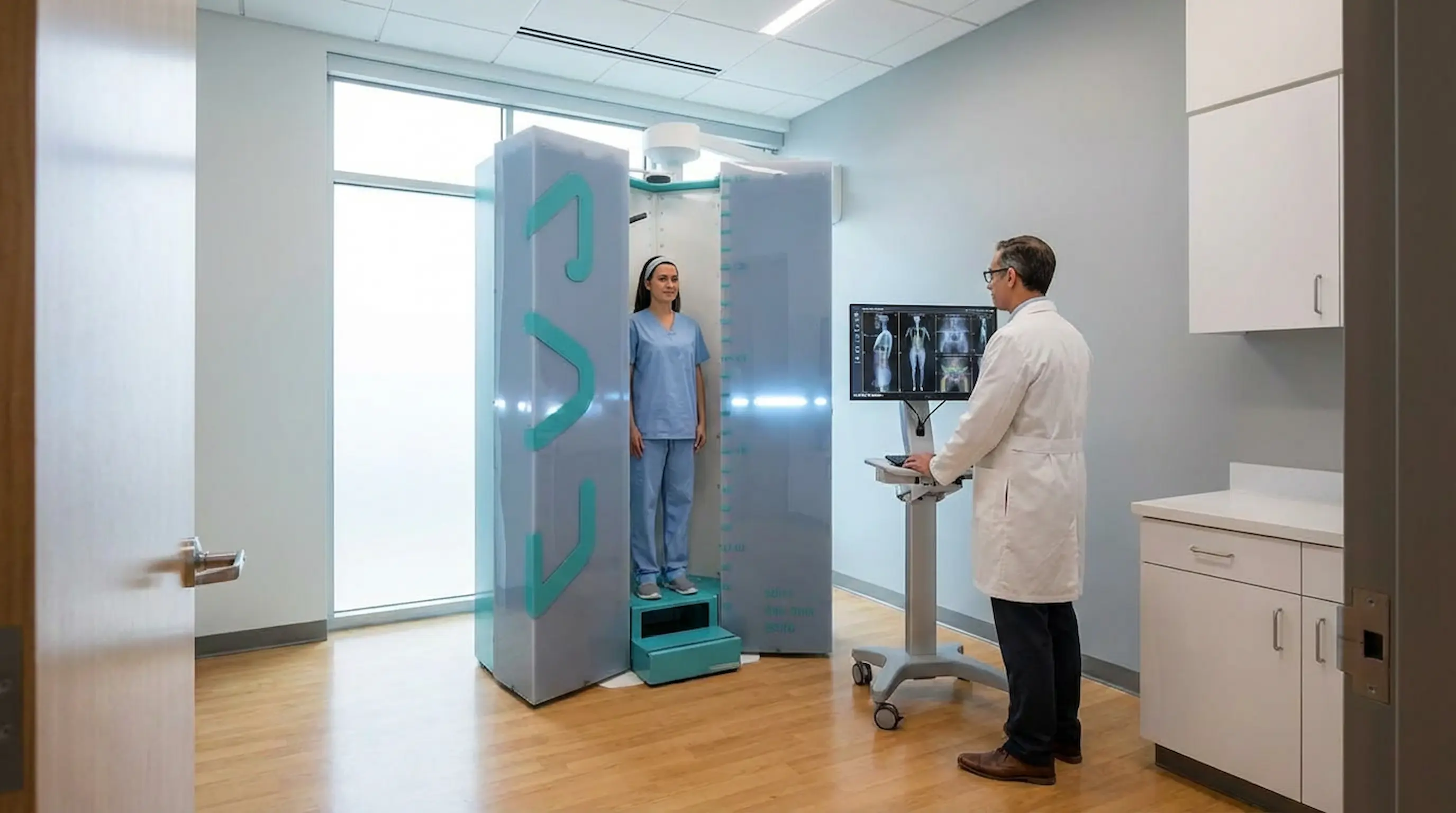
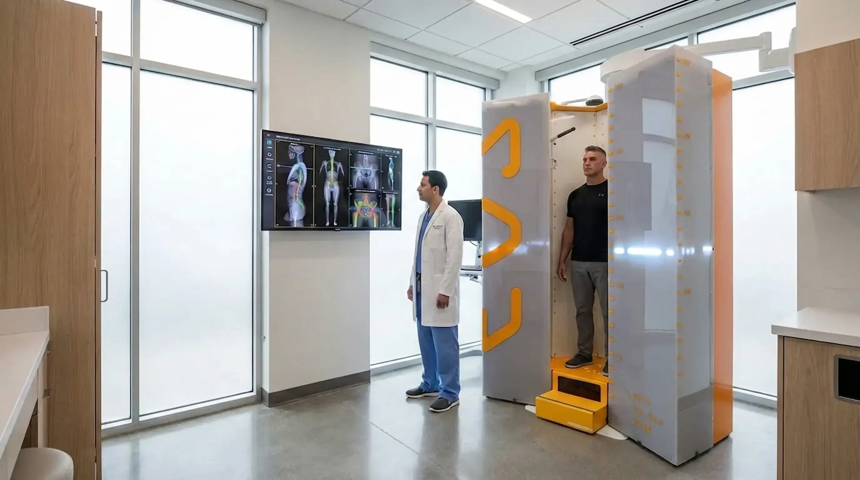
A 3D skeletal scan for sports injuries captures the full skeleton while you stand upright quite different from a standard X-ray or MRI, which are taken lying down. Lying flat removes gravity from the equation, which means the images do not show how the body truly loads during movement.
With a full-body alignment scan, clinicians can assess:
- The position and tilt of the pelvis
- Spinal curves and how they relate to the rest of the skeleton
- Knee alignment and joint angle under load
- Foot and ankle positioning
- Overall skeletal symmetry from head to feet
This level of detail is very hard to achieve through observation alone. Standing 3D imaging provides objective, repeatable measurements giving clinicians and athletes something concrete to work from rather than relying on estimation.
Musculoskeletal Imaging and the Athlete's Kinetic Chain
The body works as one connected system, often called the kinetic chain. Every athletic movement whether running, jumping, or changing direction depends on different body parts working together smoothly. When one part of this chain is out of alignment, the rest of the body begins to compensate. For example, a rotated pelvis can change knee movement, while a flat foot may affect how force travels through the hips and spine. Over time, these small adjustments create extra stress that can lead to pain, reduced performance, and repeated injuries.
Musculoskeletal imaging helps specialists see the full kinetic chain instead of focusing only on the painful area. This makes it easier to find the real source of the problem and plan better treatment or training adjustments.
Key examples of kinetic chain compensation include:
- A rotated pelvis affecting knee tracking
- Flat feet changing hip force movement
- Uneven limb loading increasing joint stress
- Poor spinal alignment reducing balance and movement control
By examining the whole body together, clinicians can better understand how alignment problems are connected and how they impact athletic performance.
Postural Asymmetry: The Hidden Factor Behind Performance Loss
Postural asymmetry means the left and right sides of the body are not evenly balanced. Some variation is normal, but when it becomes significant, it affects both performance and injury risk.
Here is how asymmetry shows up in sport:
- Balance — uneven loading makes controlled movement harder, especially in fast direction changes
- Force transfer — an asymmetrical skeleton cannot transfer power efficiently through the kinetic chain
- Movement economy — the body works harder to compensate for imbalance, creating fatigue faster
The problem is that small imbalances often feel completely normal. There is no obvious warning sign until the body is placed under repeated competition stress and by then, injury may already be developing. Catching asymmetry early, through a skeletal alignment scan for athletes, gives a real and practical advantage.
Lower Limb Alignment and Its Role in Sports Injuries
Lower limb alignment refers to the position and angle of the bones from the hip joint down through the knee, tibia, and into the foot. When this alignment is off, it changes how force travels through every step, stride, jump, and landing.
Poor lower limb alignment is linked to a wide range of common sports problems, including:
- Knee pain and patellofemoral issues
- Hip impingement and hip flexor strain
- Ankle instability and stress-related foot pain
- Altered gait patterns that reduce efficiency and increase injury risk
This matters across all sports. Runners repeat the same loading pattern hundreds of times per session small alignment errors compound quickly. Field sport athletes face sharp direction changes with high demands on the lower limb joints. In all cases, alignment affects the outcome. A skeletal alignment scan for athletes provides clear, measurable data about lower limb angles that is very difficult to assess accurately by eye.
How 3D Skeletal Scans Support Sports Biomechanics
Structure and movement are inseparable. The way the skeleton is aligned at rest directly influences how the body moves during activity and this is the foundation of sports biomechanics.
When alignment data is available, it can inform decisions across the athlete's journey:
- Training planning — coaches can programme more effectively when structural asymmetries are clearly identified
- Rehabilitation — physiotherapists can use alignment data to understand why an injury occurred and plan recovery accordingly
- Return-to-play — a structural baseline gives clinicians better confidence that the body is ready before an athlete goes back to full training
Sports medicine 3D imaging does not replace clinical judgement but it gives that judgement a much stronger, objective foundation.
Athlete Performance and Alignment: Why Structure Affects Output
An athlete performance scan is not only about preventing injury. It is also about understanding how skeletal structure may be limiting what an athlete can actually achieve.
| Aspect | Good Alignment / Performance | Poor Alignment / Performance |
|---|---|---|
| Stability | Joints load evenly, movement is controlled | Compensation patterns develop |
| Power | Force transfers efficiently | Energy lost through structural inefficiency |
| Movement Efficiency | Less effort needed per movement | Body works harder, fatigue comes earlier |
| Symmetry | Both sides contribute equally | Dominant side overloads, weaker side underperforms |
When the skeleton is well aligned, the body moves cleanly and efficiently. When alignment is off, the body adapts but those adaptations come at a cost. Performance may plateau, movements feel harder than they should, and recovery takes longer. A full-body alignment scan helps identify whether skeletal balance is contributing to these issues.
Common Athlete Problems a 3D Skeletal Scan May Help Investigate
There are a number of situations where orthopedic sports imaging may provide useful information:
- Recurrent knee pain without a clear structural cause on standard imaging
- Hip imbalance — one hip sitting higher or rotating differently from the other
- Leg length discrepancy — affecting gait and how load is distributed
- Spinal loading and posture issues — especially in athletes who lift or perform overhead movements
- Uneven weight distribution — where the body consistently favours one side
- Persistent discomfort— that worsens during standing, walking, running, or training
A skeletal scan does not diagnose injuries. It provides structural and alignment information that supports clinical conversations and guides the next steps. It is a useful tool not a standalone answer.
Who Can Benefit from a Sports Performance 3D Skeletal Scan
A sports performance 3D skeletal scan is not reserved for professional athletes. It is relevant for a much wider range of people:
- Competitive athletes looking to understand their body at a structural level
- Amateur athletes who train regularly and want to reduce injury risk
- Runners dealing with recurring lower limb problems that have not resolved
- Teenage athletes in growth phases, where alignment can shift quickly
- Athletes returning from injury who want to confirm they are structurally ready
- Anyone with persistent niggles that come and go but never fully clear up
If something has never felt quite right even without a clear diagnosis checking your eligibility for a scan at ScanAlign is a straightforward first step.
What to Expect from an Athlete Alignment Scan
The process is simple and quick. Here is how it works:
- Before the scan — no special preparation needed. Arrive in comfortable clothing with no changes to your normal routine.
- During the scan — you stand in a natural upright position inside the EOS scanner. The scan takes only a few seconds with no lying down or uncomfortable positioning required.
- After the scan — the images show the full skeleton in a weight-bearing position. These are reviewed for alignment, symmetry, and lower limb mechanics, and the results support conversations with your clinician or physio.
If you have questions before booking, the patient support page at ScanAlign covers common queries and what to expect from the process.
When an Athlete Should Consider This Type of Imaging
There is no single trigger for when to book an alignment scan. But these situations are common reasons athletes and clinicians choose this type of imaging:
- Ongoing pain without a clear explanation from previous assessments
- Repeated injuries on the same side of the body
- A visible difference in posture or movement between left and right
- Questions about lower limb alignment or leg length
- Before or after rehabilitation, to understand structural changes
- Any situation where a structural view would add useful clinical context
How ScanAlign's EOS Imaging Fits This Need
ScanAlign provides standing EOS imaging a low-radiation, full-body skeletal scan taken in a natural upright position. EOS captures the entire skeleton in a single scan while you stand naturally, giving a complete picture of the body under real gravitational load. It uses significantly less radiation than a conventional X-ray something explored in more detail in this guide on whether EOS scanning is safe.
What makes this particularly well-suited for athletes is the combination of standing position, full-body coverage, and low radiation dose. The images support conversations with physiotherapists, sports medicine professionals, and orthopaedic specialists giving those discussions a clear, objective foundation rather than relying on observation alone.
Conclusion
Better performance and fewer injuries often begin with a better understanding of how the body is built and aligned. Training harder is not always the answer sometimes the body needs to be understood before it can be improved.
Musculoskeletal imaging, injury risk assessment, postural asymmetry analysis, and lower limb alignment data all work together to build that understanding. With standing 3D imaging, athletes now have access to a tool that captures this in a clinically meaningful, low-radiation, and practical way. For any athlete who wants to take injury prevention seriously a full-body standing skeletal scan is a logical and valuable step forward.
Take the Next Step in Injury Prevention
Better performance begins with better understanding of your skeletal alignment. A standing 3D scan offers precise insight to support safer training and recovery decisions.
Book Your Appointment!FAQs
- 1. What is musculoskeletal imaging in sports medicine? It refers to imaging tools used to assess bones, joints, and skeletal structures. In sport, it helps identify alignment issues and injury risk factors that a standard clinical check may not reveal.
- 2. How can a 3D skeletal scan help with injury risk assessment? It provides objective measurements of alignment and symmetry across the full body, helping clinicians spot areas under unusual stress before they develop into a serious injury.
- 3. What is postural asymmetry and why does it matter? It means the left and right sides of the body are structurally uneven. In athletes, this can reduce power, balance, and movement efficiency and may increase injury risk if left unaddressed.
- 4. Why is lower limb alignment important for athletes? The angles of the hips, knees, ankles, and feet determine how force moves through the body. Poor alignment puts extra stress on specific joints, which leads to pain and injury over time.
- 5. Is a standing EOS scan better than a standard X-ray for alignment? For alignment questions, yes. A lying-down X-ray does not capture how the body loads under gravity. A standing scan shows the skeleton in its real functional position and uses less radiation too.
- 6. Does a skeletal alignment scan replace MRI? No. EOS scans assess bone alignment and skeletal structure. MRI is better for soft tissue detail like ligaments and cartilage. Both may be recommended depending on the clinical picture.
Continue reading
More articles
Is EOS Scan Safe? Radiation Levels, Risks, and FAQs
Discover if EOS imaging is safe, its radiation levels, risks, and benefits. Learn why it’s preferred for accurate, low-dose skeletal scans.

EOS Scan for Scoliosis: Why Standing Imaging Matters
Discover how EOS for scoliosis provides low-radiation imaging for accurate spinal alignment and better scoliosis diagnosis and monitoring.
Hip Pain & Alignment: EOS Imaging for Better Diagnosis
EOS imaging for hip pain reveals pelvic tilt, leg length discrepancy & spinopelvic alignment that routine scans miss. Book Appointment now.