
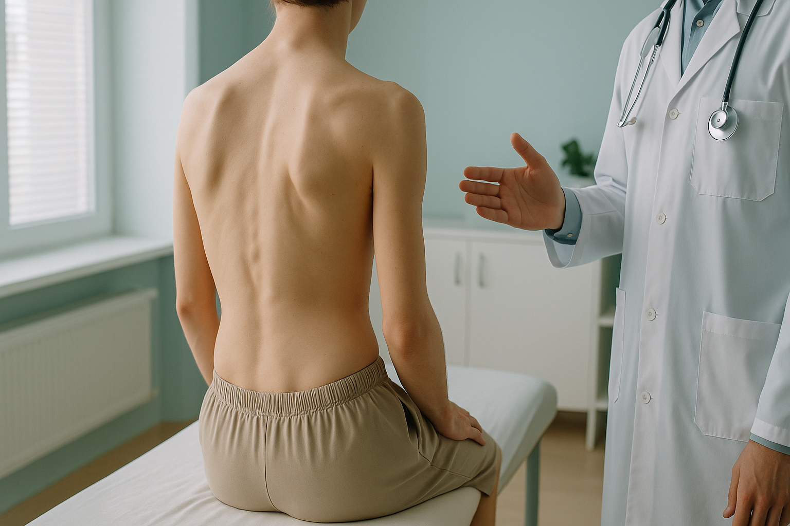
Rib Hump and Trunk Asymmetry: What Does It Signal About Your Spine?
A rib hump is a visible bulge on one side of your back, usually most noticeable when you bend forward. It happens because the spine has rotated, pushing the ribs outward on one side. It is one of the most reliable surface signs of scoliosis and means the curve is three-dimensional, not just a side-to-side bend.
Key takeaways
- A rib hump is caused by spinal rotation, not by a rib problem itself.
- It is a strong indicator of structural scoliosis in adults.
- Lying-down scans can relax the rotation and understate the problem.
- Standing, weight-bearing imaging shows the true extent of the curve and its effect on your whole alignment.
- Understanding the full picture helps your specialist plan the right approach.
What is a rib hump and why does it appear?
A rib hump is a raised area on one side of your upper or mid back where the ribs stick out more than the other side. It appears because the vertebrae in your spine have rotated along their axis. When a vertebra twists, the ribs attached to it follow, pushing backward on one side and flattening on the other.
This rotation is the hallmark of a three-dimensional spinal curve. A spine that simply bends to one side without rotating rarely produces a visible rib hump. When the hump is present, it tells clinicians that the scoliosis involves all three planes of movement: side-to-side, front-to-back, and rotational.

Many adults have lived with a mild rib hump for years without realising what it means. You might notice one side of your rib cage sticks out more when you look in a mirror, or a tailor may point out that clothes hang unevenly. These are clues worth investigating.
How do you check for a rib hump at home?
The simplest test is the Adam's forward bend test. Stand with your feet together, knees straight, and slowly bend forward at the waist with your arms hanging down. Ask someone to look at your back from behind. If one side of the rib area rises higher than the other, that is a rib hump.
One side of your mid or upper back is clearly higher than the other in a forward bend.
Shirts or jackets sit differently on each side, or a waistband tilts to one side.
Tightness or aching along one side of the rib cage, especially after sitting for a long time.
One shoulder sits higher or one hip appears more prominent, suggesting a compensatory pattern.
A home check can raise a question, but it cannot measure how much rotation is present or whether the rest of your spine and pelvis are compensating. That requires proper imaging.
What causes a rib hump in adults?
In most adults, a rib hump results from a scoliosis that developed during adolescence and was either mild enough to go unnoticed or was monitored but never treated. Over the years, degenerative changes in the discs and joints can allow the curve to progress slowly, making the rotation more noticeable.
The most common cause. A curve that began in the teens may worsen gradually over decades.
Wear on one side of the spine can introduce new rotation or worsen an existing curve.
A pelvic tilt or leg-length difference can force the spine to twist and rotate higher up.
Because the body is a connected chain from your feet to your head, a problem lower down can create a compensatory curve and rotation further up. That is why looking at only one region of the spine can miss the underlying cause.
How does standing imaging assess a rib hump?
A standing, weight-bearing image captures your skeleton in the position where gravity and your muscles are both at work. This is exactly when spinal rotation and rib prominence are at their most apparent. When you lie down for a conventional scan, your muscles relax and the curve can partially straighten, making the rotation look less significant than it truly is.

An EOS scan captures your whole body, from head to feet, in a single standing image. This means your consultant can see not only the rib hump and the spinal curve behind it, but also whether your pelvis is tilted, whether one leg is effectively shorter, and how your hips and knees are aligning to compensate. It uses about 90% less radiation than a standard X-ray, around ten times less, so it is a practical choice for ongoing monitoring.
Want to understand what your rib hump means for your whole alignment? Talk to our team about what standing imaging can show.
Book a free video consultationCan a rib hump be reduced without surgery?
It depends on the cause and severity. In some adults, targeted physiotherapy and exercises focusing on spinal rotation and core stability can improve the appearance and symptoms of a rib hump. Specialist scoliosis physiotherapy programmes, such as Schroth-based methods, are specifically designed to address rotational curves.
However, if the rotation is caused by significant structural changes in the vertebrae or discs, exercises may manage symptoms and slow progression but are unlikely to fully remove the hump. In more severe cases, a spinal surgeon may discuss surgical options.
The key point is that the right approach depends on understanding the full picture first. Without seeing how your spine, pelvis and legs interact under load, any plan is based on incomplete information. That is where whole-body standing imaging adds real value.
When is MRI more useful? EOS shows bones, joints and alignment beautifully. But if your clinician suspects nerve compression, disc problems or soft-tissue causes behind your symptoms, MRI is usually the better tool for those structures. The two tests answer different questions and often work best together.
Frequently asked questions about rib humps
Not exactly. A rib hump is a surface sign caused by the rotational component of scoliosis. You can have a mild scoliosis without a noticeable rib hump, but a visible rib hump almost always indicates that spinal rotation is present as part of a scoliotic curve.
Yes. In adults, degenerative changes in the discs and joints can allow a curve to progress slowly, which may increase the rotation and make the rib hump more prominent over the years. Monitoring is important.
The hump itself is not always painful, but the underlying spinal rotation and the compensatory muscle imbalances it creates can cause back stiffness, one-sided aching and fatigue. Pain often appears away from the hump because other areas of the body are working harder to compensate.
Specialist exercises, particularly scoliosis-specific physiotherapy, can help reduce the appearance and manage symptoms in some cases. They are most effective when guided by a clear understanding of the curve and rotation from imaging. They may not fully remove a structural hump.
This is almost always because the vertebrae in that region have rotated, pushing the attached ribs backward on one side. It is the three-dimensional nature of scoliosis that creates this unevenness, not a problem with the ribs themselves.
No. ScanAlign is a self-pay clinic and you can refer yourself directly. You start with a free video consultation to discuss whether a scan is appropriate for your situation.
Yes. The EOS system uses about 90% less radiation than a standard X-ray, around ten times less. You simply stand still for a few seconds. There is no enclosed tube or loud noise. For more details, visit the safety and FAQs page.
When you lie down, gravity is removed and your muscles relax. This can partially straighten the curve and reduce the visible rotation, making the problem look milder than it actually is when you are standing and going about your day. A standing scan captures what your body truly does under load.
For current pricing, please visit the FAQs and cost page.
Yes. Many adults with a mild scoliosis were never screened as adolescents, or their curve was too small to notice at the time. As degenerative changes develop later in life, the rotation can increase and the rib hump may become more visible for the first time.
See your whole alignment, standing up
A rib hump is a clue. A whole-body standing scan shows the full story, from your spine to your pelvis, hips and legs.
Book a free video consultation Learn about EOS imagingWritten by Abbas Dhami (Specialist Diagnostic Radiographer)
Sources
This page is general information and not a diagnosis.
ScanAlign is the trading name of CoreMed Solutions Ltd and operates under The Harley Street Hospital's CQC licence.